What is Target Engagement and how does it help my TMS?
Using rTMS in the treatment of depression, clinicians can rely on various stimulation targets. Overall the consensus is that rTMS should target the Dorsolateral Prefrontal Cortex (DLPFC), however there are several heuristics to apply this in practice, such as the use of the 5-cm. rule, the Beam-F3 method or structural MRI Neuronavigation. While all three methods aim to target the same structure, within the same individual, these heuristics could result in substantial discrepancies. On the other hand, clinical efffectiveness of these measures is rather comparable on the group level, demonstrating remission rates between 30-37% (Blumberger et al., 2019; Carpenter et al., 2012; Fitzgerald et al., 2016). Therefore, it would be valuable to have a technique available to estabish ‘target engagement’ by which one can verify that the right network is targetted, rather then relying on a heuristic based on ‘assumptions’. We'll refer to Neuro-Cardiac Guided TMS (NCG-TMS) and Heart-Brain Coupling (HBC) interchangeably in the further text.
Target engagement comprises the use of a direct functional outcome measure as a validation for targeting the correct TMS location, whereby it can be demonstrated that the right area is activated, either directly or trans-synaptically. In the same way as the motor cortex is identified by thumb movement as a demonstration of primary motor cortex activation, such functional outcome measures are thus far lacking for the prefrontal cortex and DLPFC. One proposed method is by extracting connectivity patterns to frontal areas using the sgACC as a seed region (Fox et al., 2012; Fox, Liu, & Pascual-Leone, 2013). However, such approaches require an fMRI scan from every individual patient, which is less practical in clinical practice, but above all this method has shown poor interindividual reproducibility (Ning et al., 2018), limiting its use on the individual level.
What is TMS-induced Heart-Brain Coupling?
In 2017, we published a first pilot study on a novel method that employs heart rate assessed during TMS stimuation, to verify ‘target engageent’ of the Frontal-Vagal network, that considerably overlaps with the depression network (see figure below from Iseger et al., 2019) which we initially termed Neuro-Cardiac Guided TMS or NCG-TMS. More recently we advanced this method to TMS induced Heart-Brain Coupling (HBC), which is a novel iteration over NCG-TMS and is more reliable and robust on the individual level. Since this early observation in 2017, this finding has been independently replicated by several other labs.
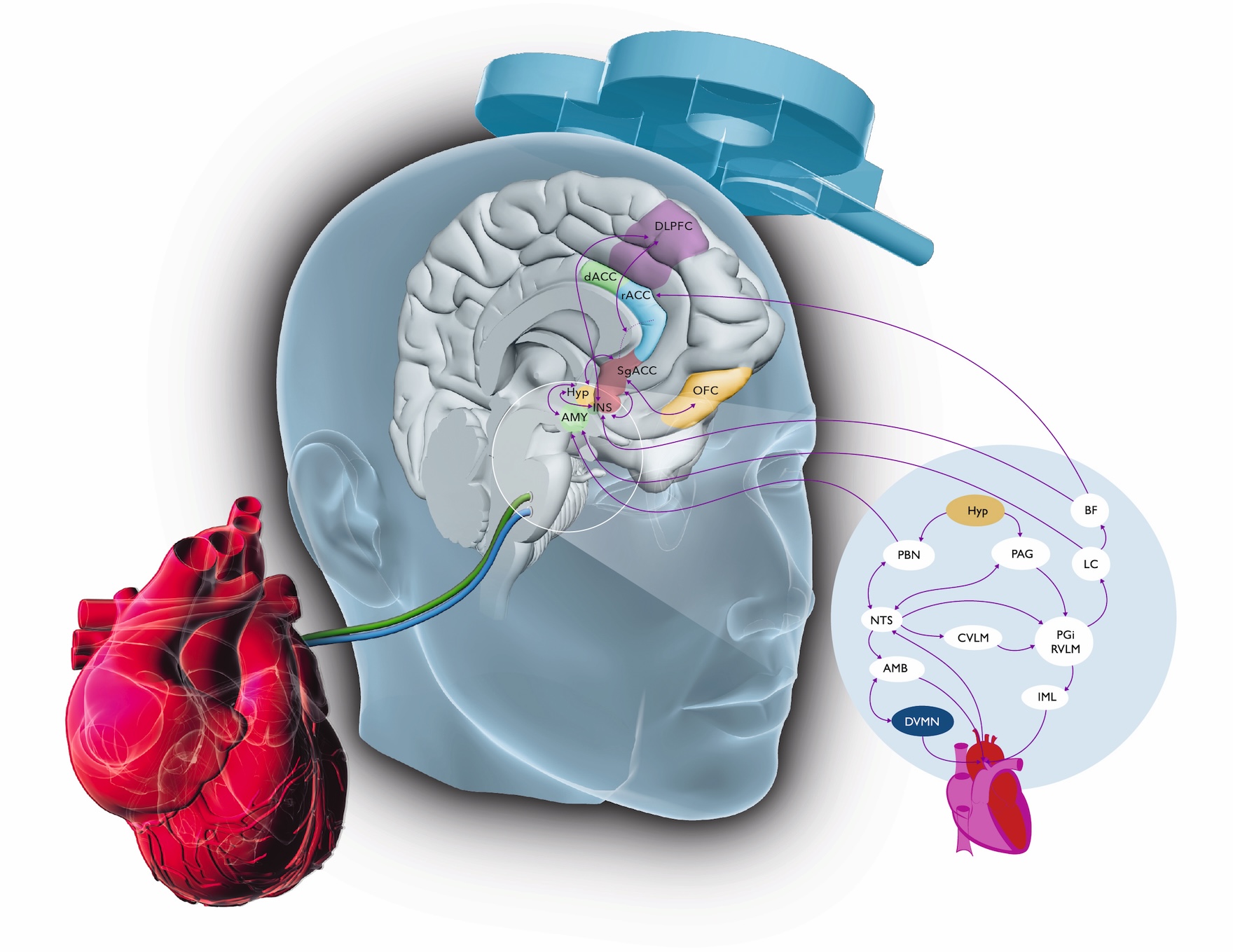
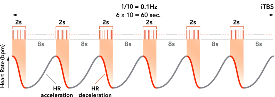
The depression network and the brain-heart axis (regulated by the Frontal-Vagal pathway, depicted above) are interconnected and overlap. Stimulation of the DLPFC has been shown to reduce heart rate (Makovac et al., 2016). The parasympathetic effects on the heart are short-lived; stimulation of the vagus nerve therefore usually results in an immediate response of the heart, typically occurring within the cardiac cycle in which the stimulation occurred, with a peak in heart rate deceleration within 5 seconds (Buschman et al., 2006). The return to a normal HR is very quick after the activity of the vagus nerve is reduced (Shaffer, McCraty, & Zerr, 2014). Heart-Brain Coupling thus depends on the stimulation duty-cycle applied, also see figure below. Here a standard ‘iTBS’ like stimulation is visualized with 2-s stimulation ‘On’, resulting in a HR decrease, whereas in the intertrain interval of 8-s HR normalizes again to baseline, where after 8-s the next train of stimulation occurs decreasing HR again. This patterned stimulation thus results in an entrainment frequency of 1/[duty-cycle]=1/10=0.1Hz. Frequency specificity of this method has also been shown for other stimulation patterns for instance with a 16-s duty cycle, indeed a 1/6=0.625 Hz HR entrainment is observed (Dijkstra et al., 2023).
Heart-Brain Coupling TMS validation studies
Ever since the introduction of HBC as a target engagement technique, one of the main premises was that TMS induced Heart-Brain Coupling would be indicative of transsynaptic actvation of the anterior cingulate cortex (ACC), more specifically the subgenual ACC. In a recent fMRI validation study in collaboration with Shan Siddiqi we were able to confirm this, where fourteen people were stimulated under fMRI based neuronavigation, to their individual prefrontal site that was a) anticorrelated to the sgACC (meaning it is functionally connected to the sgACC); b) positively connected or, c) non-correlated to the sgACC. When only inspecting HBC from the Heart-Brain Connect iPhone app as an agile readout, we could succesfully identify an sgACC-anticorrelated site in 12/14 cases - or a 86% accuracy (Dijkstra et al., 2024), demonstrating sites with strongest HBC indeed are sites functionally connected to the sgACC.
In a further validation work, colleagues from the US and Canada shared heart-rate data with us from controlled TMS studies that included both sham and real TMS stimulation. When we analysed these datasets blinded to group assignment we succesfully 'unblinded' the CARTBIND study with 83% and the SAINT data with 90% accuracy, demonstrating how clearly different HBC is between real and sham stimulation with a large (ES=1.4; Dijkstra et al., 2023). Furthermore, we here also found the prefrontal site that elicited strongest HBC, varied considerably across participants and in recent work demonstrated responders to TMS demonstrated significantly stronger HBC (Dijkstra et al., 2025) confirming utility of HBC to improve clinical outcomes.
Further reading and HBC software
We are currently finalising our new Heart-Brain Connect - Live software, that will allow real-time determination of the best prefrontal HBC sites, stay tuned... for now, the Heart-Brain Connect app is still available for iPhone.
The Heart-Brain Coupling app for iOS
The Brainclinics Foundation developed Heart-Brain Connect, a breakthrough Heart-Brain Coupling app to assist clinicians in pinpointing the optimal location for rTMS treatment. Our unique approach has great advantages over more traditional methods and can be tried for free. The app can also be used as a resonant breathing training method.
Heart-Brain Connect is available in the iOS App store.
PhD theses and literature
Several full text PhD theses on this topic from Brainclinics alumni can be found under Brainclinics Insights and our scientific publications can be found under publications.